
Nuclear mechanics as a diagnostic and therapeutic target for glioblastoma
Glioblastomas (GBMs) are the most lethal primary brain tumours. The absence of effective therapies is mainly due to tumour invasion and to the resistance of invading cells to treatments such as radio and chemo-therapies. In GBMs, lamin proteins that control nuclear envelope stiffness, have recently emerged as potential markers of aggressiveness and tumourigenicity. Nuclear mechanics has appeared as a key determinant of cancer cell invasion leading us to hypothesize that genes controlling nuclear mechanics of GBM cells may be used as diagnostic tools and potential therapeutic targets to improve the prognostic of GBMs.
The working hypotheses of this M2 internship project is that alterations in nuclear mechanics contribute to GBM aggressiveness and directly influence cell invasive behaviour. The intern will first use clinically annotated primary patient-derived GBM cells and rheological techniques (optical tweezers, microfluidics) to measure nuclear morphology and mechanics (Figure). Second, he/she will modulate the expression levels of lamins to modify both nuclear mechanics and GBM cell invasion and test whether lamins could be used as potential molecular targets to control GBM aggressiveness.
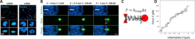
Figure: A. Comparison of the morphology of the nucleus in two different GBM cell lines (U3039 and U 3031). B. Measurements of the viscoelasticity of the nucleus using indentation of GBM nuclei in living cells. A. Images showing a typical nuclear indentation experiment. The white cross represents the centre of the optical tweezers in which the 2 µm- diameter bead is trapped (green). The nucleus (blue) is indented by moving the cell towards the right (white arrow) which displaces the bead away from the trap centre of a distance ∆x. C. Scheme of the bead in the optical trap. D. Force-indentation curve showing the force F as a function of the indentation d in the experiment shown in B.
Key words: nuclear envelope; lamin A/C; lamin B1; lamin B2; LINC complex; optical tweezers; microfluidic; cancer; glioblastoma; cytoskeleton; migration; invasion.
Collaborators: Sandrine Etienne-Manneville (Institut Pasteur, Paris), Catherine Villard (Institut Curie, Paris), Wang Xi (IJM, Paris), Nicolas Borghi (IJM, Paris)
Laboratory: Matière et Systèmes Complexes, UMR 7057 CNRS-Université de Paris, 10 Rue Alice Domon et Léonie Duquet, 75013 Paris
Contact: Jean-Baptiste Manneville (Jean-Baptiste.Manneville@u-paris.fr)
À lire aussi

spectroscopie de plasma induit par laser pour analyser l’adsorption par immersion d’ions métalliques par des hydrogels d’alginate séchés
Enrique Manso et Alain Ponton du laboratoire MSC, en partenariat avec des chercheurs de l'université de la Corogne (Maria Pas Mateo, Gines Nicolas) en Espagne, présentent une application originale de la spectroscopie de plasma induit par laser (LIBS) pour analyser...

Poste Technicien.ne en Biologie
Un poste (CDI) de Technicien.ne en Biologie est ouvert au concours externe Ingénieur/Technicien (IT) du CNRS pour rejoindre le laboratoire Matière et Systèmes Complexes (MSC UMR 7057 CNRS - Université Paris Cité) à Paris. Lieu : Laboratoire MSC, Paris Profil :...
Le youtubeur MonsieurLeChat au labo
Et si le caramel permettait de comprendre l’érosion d’un paysage ?Michaël Berhanu a reçu dans son laboratoire le youtubeur Monsieur Lechat, pour lui parler de dissolution. La vidéo est ici.
Face à la rigidification des sols, les racines développent une capacité d’auto-adaptation inédite
Une équipe du laboratoire Matière et Systèmes Complexes (MSC – Université Paris Cité/CNRS), impliquée dans une collaboration internationale, met en évidence un mécanisme original permettant aux racines de s’adapter à la rigidification des sols, un phénomène accentué...