Abstract below
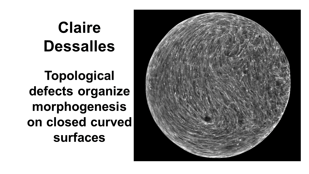
Claire Dessalles
Institut Lumière Matière, CNRS-Université Lyon 1
Topological defects organize morphogenesis on closed curved surfaces
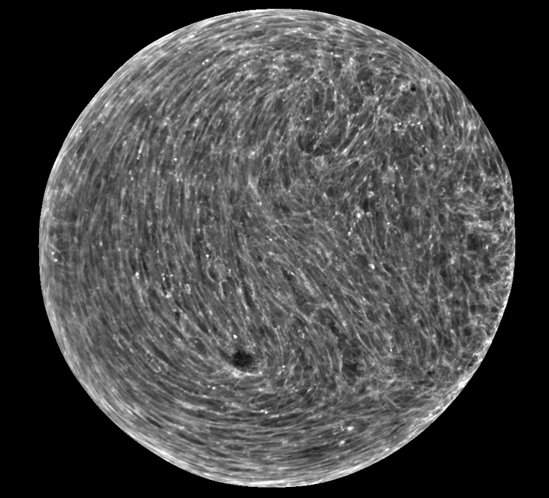
Morphogenesis, the process by which tissues acquire their shape, hinges on a finely orchestrated collective motion of cells. Accumulating evidence shows that many biological tissues behave as active nematics, both in vitro and in vivo. The collective motion of cells is controlled by the nematic order, and topological defects have been proposed as morphogenic organizers via active stresses. However, the generation and control of tissue-scale forces involved in morphogenesis remain poorly understood, in particular within 3D surfaces.
To investigate how geometry and topology control morphogenesis, I grow myoblast cells on the surface of alginate microspheres and monitor the nematic field, cellular flows, and tissue growth. When the tissue reaches confluency, four equidistant +1/2 defects are observed in the actin network, consistent with the topological charge imposed by the sphere (see image below). Subsequent growth of the monolayer due to continuous proliferation shows the formation of a multilayered tissue with two main orthogonal orientation. Upon further growth, the half defects fuse by pair, forming two +1 defects, and the thickness of the tissue becomes heterogeneous with the presence of two mounds co-localizing with the +1 defects. Together, the defect fusion and mound formation form a first spontaneous symmetry breaking event. Finally, the two +1 defects migrate towards one another and form a +2 region, accompanied by the fusion of the two mounds into one main protrusions. In this synthetic model system, a complex and spontaneous morphogenesis emerges from the interplay between the topological defects and cellular flows, illustrating the role of physical principles in a fundamental biological process.