Abstract below
PhD thesis defense of Saatvik Potluri
The characters in zebrafish larval tail fin regeneration: A complete story”,
PhD Supervised by Nadine Peyriéras.
Location: Amphitheatre 2, Olympe de Gouges building
Abstract:
Tissue regeneration is a synchronous process that involves leukocytes, fibroblasts, and extracellular matrix (ECM) components. The zebrafish larva can regenerate a functional tail fin within 3 days of amputation. The regenerative capacity of zebrafish serves as a model to study scar-free regeneration.
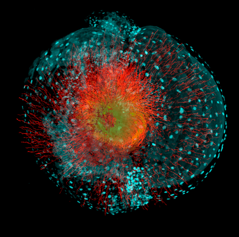
We use 3D+time imaging of transgenic reporter lines in which we label neutrophils, macrophages, and fibroblasts. We detect RNA and protein expression profiles to identify cell identity and function. We characterize the ECM composition and texture using STED microscopy. These approaches show the spatiotemporal dynamics of cells and the ECM during tail fin regeneration.
Neutrophils are recruited to the tail fin site only briefly, while macrophages are kept on site throughout regeneration. Macrophages polarize from an inflammatory to an anti-inflammatory state and secrete TGFβ. This activates resident cells to become myofibroblasts that express pSMAD3, αSMA, and PAI-1 RNA.
Cell tracking of PAI-1:GFP+ fibroblasts using a transgenic line showed a change in their behavioral dynamics. After their transient expression of PAI-1 RNA, they produced postnb RNA, an ECM component necessary for regeneration. PAI-1:GFP+ cells were not responsible for repopulating the tail fin and instead migrate to a posterior location after having secreted ECM components.
Cells in the tail fin express ECM components, such as col1a1, col12a1b, and postnb RNA. Expression of ECM components peaks at 36-48 hpa, along with P4Hβ, an ECM remodeling component.
At the time of increased ECM expression, we observed PU.1+ fibroblasts at the site of injury. PU.1 is a transcription factor typically associated with cells of hematopoietic origin. Nonetheless, we found PU.1+ fibroblasts and characterized them as a fibroblast subpopulation distinct from PAI-1:GFP+ fibroblasts. We carried out preliminary investigations on PU.1+ fibroblasts to characterize their identity and function.
Our investigation extends to the architecture of the evolving ECM. Fibronectin, col1a1 and col12a1a were initially present in punctae. They were later present as fibrillar structures at the end of regeneration. We also detect other distinct features of the ECM, such as tubular structures specific to the regenerating tissue.
Our results show the diversity of fibroblast populations in zebrafish larval tail fin regeneration. The interactions between leukocytes and resident cells initiate a sequential pattern of cell behavioral dynamics to repopulate the tail fin and remodel the ECM. We characterize the transition of the tail fin through different phases to regenerate tissue.
Keywords: Regeneration, fibrosis, fibroblasts, macrophages, extracellular matrix, TGFβ, PAI-1, PU.1, zebrafish, 3D+Time and STED microscopy.