Vertebrate organogenesis
Participants: Nicolas Chevalier, Monique Frain, Nadine Peyrieras, Thierry Savy
Organogenesis and mechanobiology of the intestine
Our work focuses on the morpho- and patho-genesis of two smooth muscle organs: the intestine and the uterus. In the intestine, we study the biophysical factors – mechanical and electrical – involved in the embryogenesis of this organ, from the enteric nervous system (the “second brain”) derived from neural crest cells, to smooth muscle and interstitial cells of Cajal (the “pacemakers” of the intestine). We are also interested in the physiology and hydrodynamics of the adult digestive tract, using murine models. With regard to the uterus, we are seeking to understand the origin of the uterine hypercontractility observed in endometriosis, a pathology that affects 10% of women of childbearing age. Contractility could be a fundamental cause of the development of this disease, and a therapeutic target. Our approach is both structural (electron microscopy, immunohistochemistry) and physiological (measurement of contractility, calcium imaging). We work in collaboration with hospital staff and biologists on both themes.
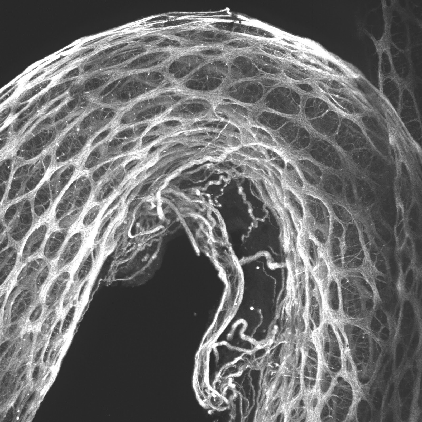
Morphogenesis and wound healing in zebrafish
The arrival of the biologists in the team is concomitant with the completion of the two theses in the ImageInLife 2017-2021 ITN focusing respectively on “multiscale dynamics in the morphogenesis of the rhombencephalon in zebrafish” (Mageshi Kamaraj, direction Monique Frain) and “Multiscale dynamics during gastrulation of the zebrafish embryo and embryoid” (Svetlana Jovanic, direction Nadine Peyriéras, co-direction Thierry Savy). Svetlana Jovanic’s thesis work could find a follow-up in the ERC synergy project HETEROMORPH led by A. Sarti and submitted in 2022.
Saatvik Potluri’s current thesis [European contract ITN INFLANET (2021-2025)] focuses on the urokinase pathway and extracellular matrix remodeling in experimental models of inflammation in zebrafish (supervisor Nadine Peyriéras, co-supervisors Monique Frain and Thierry Savy). Since arriving at MSC, the student has established a model of inflammation in larval zebrafish based on caudal fin ablation. A tool for sectioning under the microscope has been developed. The cutting operation will be robotized. Conditions for 3D+time multiphoton microscopy observation and monitoring of mobilized cells at the wound site have been established. Molecular tools for labelling the extracellular matrix and components of the Urokinase pathway have been developed and validated.
…
Read more
Internship/PhD on Hydrogels for adsorption of toxic metals
Proposal for master internship/PhD Hydrogels and composites with optimal efficiency and minimal environmental impact for adsorption of toxic metals The increasing demand for water due to technology and industrial activity has led to the use of hydrogels to treat...
Postdoc on the acoustics of membrane materials
As part of the Membranes project, the MSC laboratory is carrying out experimental research into the acoustic properties of membrane media, i.e. media whose internal structure contains thin layers of materials that can provide an original elastic response [1]. Examples...
Postdoc on heterogenous cavitation
We are looking for a post-doc to join our research team and perform experiment on heterogeneous cavitation to explore the origin of cavitation nuclei. The apparition of macroscopic bubbles inside a liquid either by boiling when temperature increases, from cavitation...
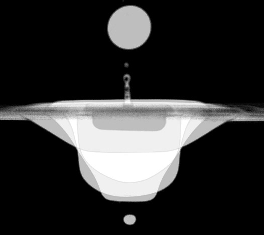
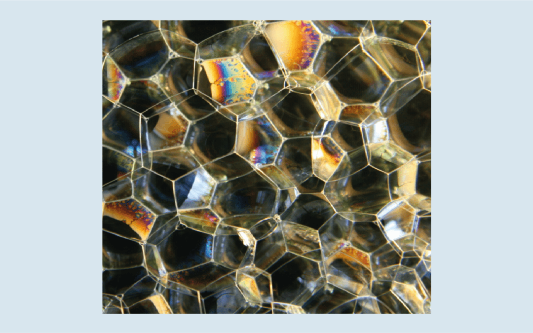
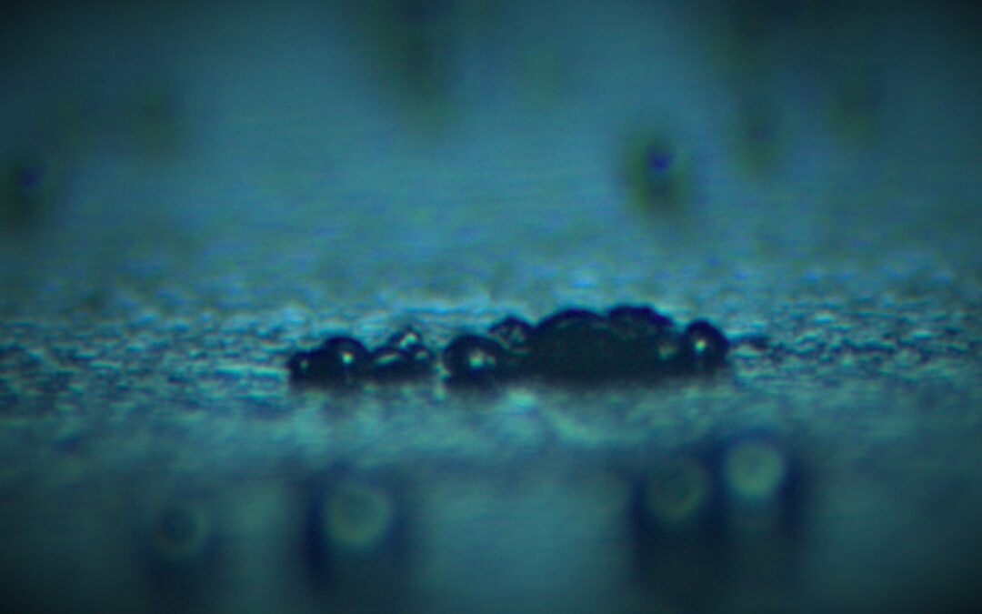
Bubble entrapment by drop impact
Only under certain conditions does a drop falling onto a bath entrap an air bubble. We propose a phenomenological law that describes these bubbling conditions in terms of Froude, Weber, and capillary numbers.Figure. Superimposed images of a drop and the cavity it...