Tuesday, November 5th, Bâtiment Condorcet, 10 rue Alice Domon et Léonie Duquet, 75013 Paris.
PhD defense of Lorijn van der Spek
Application of mechanical and geometrical constraints of muscle cells to promote differentiation at different scales
Supervised by Myriam Reffay et Sylvie Hénon (Physique du vivant, MSC)
Abstract:
This thesis explores still unknown features of the interplay between shape, mechanical stimulation and muscle cell differentiation. The muscle is a multiscale system built up of myofibers. Myofibers differentiate from individual muscle stem cells or myoblasts, in a process in which they elongate and align. It is well-known that stimulating collections of myoblasts by stretching them promotes their differentiation. This interplay between stretching and differentiation is studied in this thesis at both single-cell scale in microcontrolled 2D environment and in lab-made 3D tissue.
At single-cell scale, myoblasts have been placed on square and rectangular adhesive micropatterns to determine whether geometrical constraints result in a different differentiation fate. The degree of differentiation has been measured using different markers and techniques to quantify proliferation rate, membrane potential and the expression of differentiation markers. All these factors vary with the shape of the micropattern, showing that placing cells on rectangular micropatterns inhibits their proliferation and promotes their early differentiation.
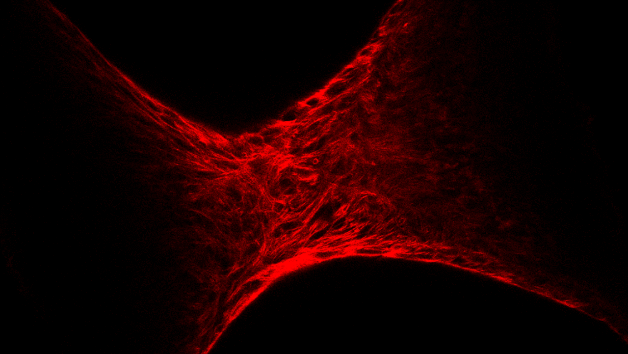
In lab-made 3D-tissue, this phenomenon was further investigated using a device developed in our laboratory: the mechanical stretcher. By magnetically labelling the myoblasts, they quickly form a much larger tissue than by using other techniques, which can also be stimulated. Pulling on this tissue causes a collective alignment of the actin cytoskeleton of the cells in the direction of the stretch. The surface phenomena of the specimens as well as local phenomena within the specimen were studied. For this purpose, a technique was developed to visualise mRNA of mature muscle differentiation markers and actin, up to 400 μm inside the tissue.
The defence will be in English.